Cornea Treatment Powered by Sharp Sight Eye Hospitals
- Delivering exceptional outcomes with state-of-the-art technology and skilled ophthalmic surgeons
All Round Cornea Treatment by Sharp Sight
- Top-Notch Eye Care Services
- Learned Eye Specialists
- Advanced & Affordable Treatments
Generally, A Corneal Transplant Is Needed In The Following Circumstances
Identify and understand the key risk factors that contribute to the development of cataracts
Ageing
Vision correction with eyeglasses or contact lenses becomes impossible
Medicines
Swelling cannot be relieved by medications/contact lenses.
Exposure
Corneal failure occurs after any other eye surgery, e.g.: cataract surgery
Chronic Disease
Due to keratoconus, a condition that causes thinning of the cornea
Trauma
There’s corneal rejection after a first corneal transplant
Habits
Scarring caused after an injury
Different types of Cornea Problems
Sharpsight Hospitals offers a comprehensive range of advanced surgical options, ensuring the highest standards of care and precision.
- CORNEA
- KERATOCONUS
- Intracorneal Ring Segments (Intacs) For KERATOCONUS
How does our Eye Function?

The rays of light from an object strike the transparent cornea and bend (refract) to enter through the pupillary aperture and pass through the lens filter to form an image on the central part of retina (macula) which acts as a film. This image signal is carried to the brain by the optic nerve which perceives it as the object known.
This is how every component of the eye is important for visual perception.
Cornea Transplant
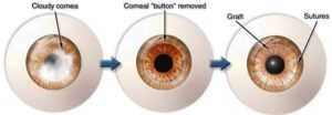
If there is any kind of obstruction at the cornea affecting light rays from entering the eyes, we go ahead with a Cornea Transplant wherein we remove the diseased cornea and replace it with a donor cornea obtained from a cadaver(within 6 hrs. of death)

Why Is Cornea Transplanted?


There are a numerous reasons why we need to change the Cornea, if you are be suffering from any of the following:
- Corneal Scar
- Corneal Ulcer
- Corneal Dystrophy
- Pseudophakic Bullous Keraopathy
- Severe Spheroidal Degeneration
- Advanced Keratoconus with apical scarring
- Trauma
Who Can Undergo Cornea Transplantation?
For a favorable outcome following are the prerequisites:
- Perception of Light must be present
- Projection of rays accurate in all quadrants
- Intraocular pressure between 10-25mm of Hg
- No Retinal or Choroidal detachment in posterior segment
- Lids and Adnexa should be healthy
- Regular follow-up be ensured
- Compliance to medication at given intervals to be strictly observed.
- High risk of graft rejection/failure should be acceptable depending on host factors
How Is Cornea Transplantation Done?

What Are The Different Types Of Cornea Transplant?

Full Thickness Penetrating Keratoplasty (PKP)
Wherein all the layers of diseased cornea are removed and replaced by full thickness donor corneal graft
Deep Anterior Lamellar Keratoplasty (DALK)
Wherein all the layers of diseased cornea are removed and replaced by full thickness donor corneal graft
Keratoconus is a progressive condition of eyes which is caused when the otherwise round cornea bulges out into an irregular cone due to its excessive thinning. It is the conical shape in a Keratoconus-affected eye that causes distorted vision, primarily because it restricts the light (entering into our eyes) from being focused correctly on the retina. Therefore, in a Keratoconus-affected cornea, light rays enter the eye at different angles instead of one focused point. However, in its earlier stages, the disorder cause blurred or distorted vision which progress if left untreated. As Keratoconus progresses, it affects both the eyes differently and its first signs are usually felt in mid-to-late teens. However, with the progression of the condition, the cornea ends up bulging out more thereby, affecting vision severely. In some cases, the cornea may swell and cause a sudden & significant loss of vision.
Soft contact lenses are generally prescribed to correct mild vision changes caused due to Keratoconus. Later, as the disorder progresses, and cornea continues to become conical, rigid gas permeable contact lenses are prescribed to be used. However, to maintain healthy eyesight and arrest the progression of Keratoconus, it is important to get your eyes checked regularly.
Keratoconus Treatment
Keratoconus can be diagnosed clinically by Slit Lamp Examination and it can be confirmed by Corneal Topography test
Spectacles and contact lenses:
Contact lenses are required for the visual improvement in patients with keratoconus. Various contact lens options, such as rigid gas permeable (RGP) lenses, soft and soft toric lenses, piggy back contact lenses (PBCL), hybrid lenses and scleral lenses are available. Use of contact lenses ensures better best corrected visual acuity in these highly irregular corneas.
Corneal Collagen Cross Linking
Keratoconus is first managed by Corneal Collagen Cross-Linking or CXL technique, which is a new modality of treatment that has a higher success rate in arresting the progession of the condition. In most cases, corneal Cross-Linking reduces the need for a corneal transplant. The treatment involves removing the superficial layer (epithelium) from the surface of the cornea and then applying Riboflavin eye drops to the eye. The eye is then exposed to UVA light. After the treatment, a bandage contact lens is worn for 1-3 days until the surface of the eye has healed. Antibiotic and steroid eye drops are also prescribed for a few weeks. This has been shown in studies published worldwide over a long follow-up period, to be successful in arresting the progression of the condition in most cases
At Sharp Sight we have the latest technology for performing the procedure which reduces the time needed on table by using an accelerated protocol.
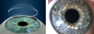
Intacs or Intracorneal rings are thin, semi-circular transparent inserts which are designed to be placed surgically in the affected cornea. This helps in reshaping or flattening the bulging cornea. These corneal implants are an effective treatment option for Keratoconus patients as it improves their best corrected vision. Intacs were approved under a Humanitarian Device Exemption (HDE) by the FDA in July 2004, allowing INTACS to be used for treating keratoconus.
Benefits of Intacs
- Requires no post-surgery maintenance
- Intacs can be removed/replaced depending on the patient’s convenience
- Safe, quick and predictable procedure
- Improvement in best glasses/contact lens corrected vision
Why Choose SharpSight FOR KERATOCONUS
At Sharp Sight Eye Hospitals we offer treatment of Opaque Cornea or Corneal Blindness through Cornea Transplant. We have a team of Keratoconus specialist who excel in cornea transplantation surgery where a healthy cornea is transplanted in place of a diseased cornea. We offer the best Keratoconus treatment through the latest equipment and world-class infrastructure. Our Keratoconus treatment cost is affordable. We pride ourselves in being among the top ten eye hospitals to offer the best Keratoconus treatment in India.
Why Sharpsight forCornea Treatment?
We've a strong portfolio of Happy Patients
We have served more than a million patients and have helped them see this world clear.


